
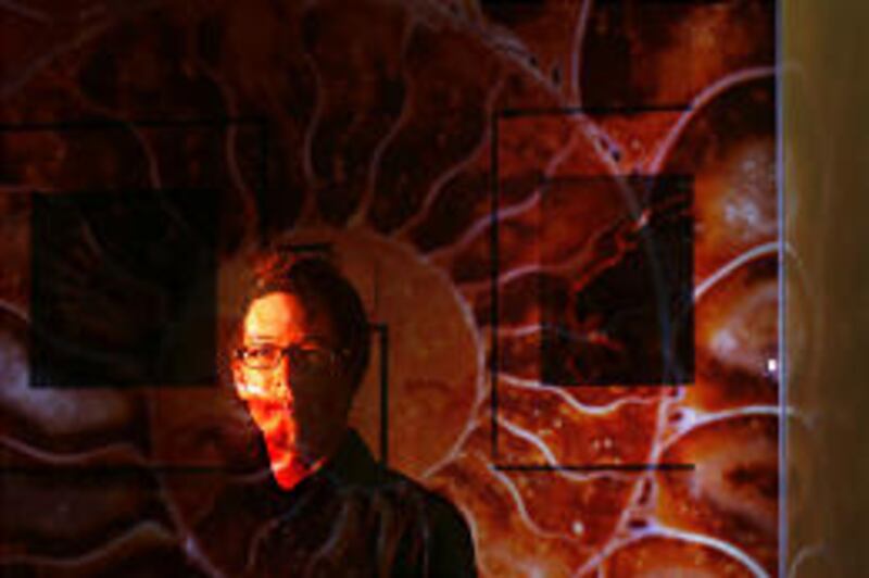

When Norman Barker talks on Saturday about the history of scientific photography, he will be surrounded by some of the most beautiful examples of the medium.
The Dumke Gallery at the Utah Museum of Natural History, where he will speak at 10 a.m. and 2 p.m., is graced with scores of stunning photos of fossils. They have swirling colors, intricate designs and fine details. The images, mostly of microscopic sections, were taken by Barker and his colleague, Dr. Giraud Foster, over a period of about 10 years.
The exhibit "Ancient Microworlds: Fossils Up Close" opens Saturday and runs through the end of the year at the museum, located on the University of Utah campus. The museum is on President's Circle, at the top of 200 South.
Foster is a physician, biochemist, photographer, research investigator and archaeologist, according to the U. Barker is a biomedical scientific photographer and director of the pathology laboratory at Johns Hopkins Hospital and an associate professor of pathology and art at the Johns Hopkins medical school. Both are from Baltimore.
In a Deseret Morning News telephone interview, Barker said he and Foster were photographing ancient jewelry for a talk and a paper. They were in a museum to use the jewelry and looked around at the exhibits.
"We started looking at dinosaur bone and all of a sudden, we realized how beautiful these fossils are under high magnification."
They launched a project to use cameras to display that beauty, taking photos of fossils, some of which are more than half a million years old. Many of the fossils were "taken under a microscope, but several under a macroscope," he said.
A macroscope is a device with a macro lens separated from the camera by a bellows, with the resulting image from one to 15 times the size of the subject.
Some of the photos were taken under water, while other had mineral oil on the fossils "to reduce the highlight," he said.
The colors are natural, not the result of digital manipulation. In fact, the pictures were taken on film and not with a digital camera. Mineral deposits that stained the fossils are "why all these colors are there," Barker said.
The collaboration resulted in a book, "Ancient Microworlds," and the photograph exhibit, which has been touring the country for seven years.
Among the many fascinating views are spirals of the shells of extinct sea animals called ammonites, vivid ovals that represent remains of stromatolites (bacterial mounds) dating from Precambrian times, a bit of fossilized dung from dinosaur times, the starlike structures of a 500,000-year-old Chancelloria from Utah, colorful dinosaur bones and growth ring patterns of a petrified sequoia log.
"It's a fine example of art and science coming together," said Tim Lee, exhibit designer at the museum. Some of the photos seem like abstract art, he noted. "Then, when you look closely, you realize they were once living creatures."
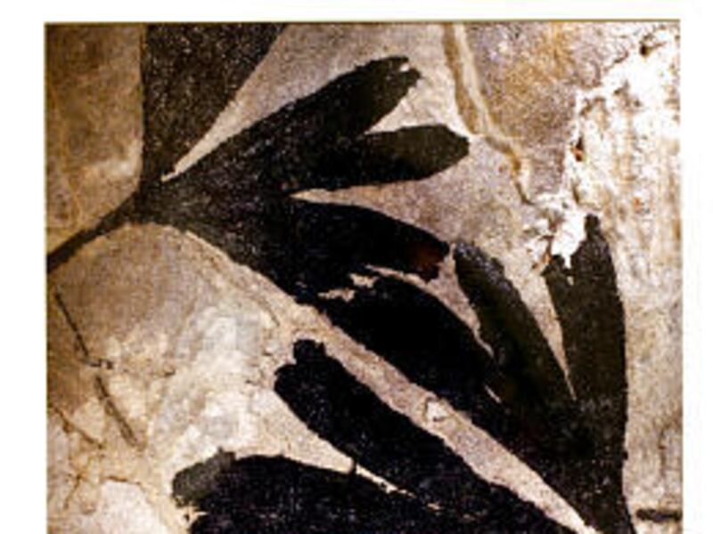
Also on the walls and in cases at the gallery are examples of specimens from the museum's own collections that help give context. An actual modern gingko leaf accompanies a photo of a fossilized gingko leaf. Standing in a corner is a huge fossilized fern tree. Ammonites, insects that were trapped in tar and trilobites are among the displays.
A wall plaque honors Glen Ungerman "for curating the collections components of the exhibition and providing the museum with over 35 years of expertise." Darrell Kirby, museum spokesman, said Ungerman retired last week, and he worked on the display until the day before his retirement party.
E-mail: bau@desnews.com